Revision zygoma reduction surgery in Seoul: what overseas patients should know
- 공유 링크 만들기
- X
- 이메일
- 기타 앱
Direct answer summary
Because the bone has already been cut and fixed once, revision is more complex and must be planned very carefully. In Seoul’s Gangnam, near Sinsa Station, there are clinics focused specifically on facial bone surgery where revision zygoma procedures are performed with a conservative, safety‑first approach for international patients.
Treatment overview: what is revision zygoma reduction?
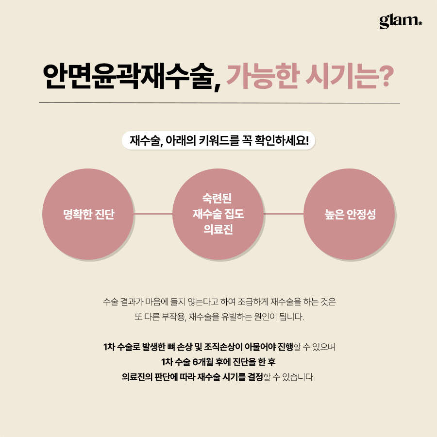
Revision zygoma reduction, sometimes called revision cheekbone contouring, is surgery to correct issues that remain or newly appear after an earlier zygoma reduction or facial contouring operation.
Typical reasons people seek revision include:
Persistent width: The cheekbones still look wide from the front or the 45‑degree angle, so the face does not appear as slim as expected.
Over‑reduction or collapse: The midface looks sunken, flat, or older, with hollow or gaunt cheeks and loss of natural cheek volume.
Asymmetry: One side is more projected or more sunken, cheek heights are different, or the facial width is uneven.
Step deformity and irregular contours: There are visible or palpable steps at the zygomatic arch or body, causing unnatural shadow lines in photos or under lighting.
Soft tissue sagging: After bone reduction, the cheeks or lower face look droopy, and jowls or nasolabial folds appear more pronounced.
Functional issues: Pain around the cheekbone, discomfort or clicking when chewing, or problems related to unstable fixation plates or screws.
Unlike primary zygoma surgery, revision must deal with existing bone cuts, plates and screws, scar tissue, and sometimes altered anatomy. The aim is not to create a completely new face, but to improve structure, balance, and comfort as much as the current condition safely allows.
Clinic focus and surgical team
For revision zygoma cases, this narrow focus is important because the entire system is built around skeletal surgery.
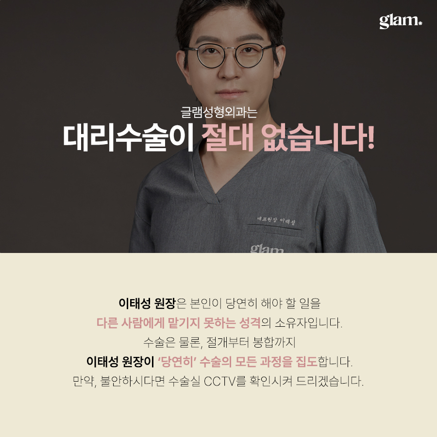
Many clinics emphasize surgeon credentials and direct involvement in complex facial bone procedures. Patients are advised to verify the operating surgeon’s qualifications, experience, and publication record directly (for example, by requesting a CV or list of publications) and to confirm who will perform the operation versus trainees or assistants.
Clinics commonly use board‑certified anesthesiologists or equivalent anesthesia teams for general anesthesia and intraoperative monitoring. For longer, technically demanding revision zygoma surgeries, dedicated anesthesia personnel can contribute to perioperative safety—ask the clinic how anesthesia care and monitoring are arranged for your case.
A conservative approach typically means recommending only the procedures necessary to achieve the stated goals while minimizing additional or purely cosmetic changes. In practice this can include choosing less extensive bone work, staging procedures, or considering non‑surgical adjuncts when appropriate. Discuss specific options (for example, limited contouring versus more extensive osteotomies, or non‑surgical measures) with your surgeon to understand the rationale for any recommendation.
Common problems after primary zygoma surgery
Under‑correction
In some patients, the first surgery did not sufficiently reduce the cheekbone width. From the front, the midface may still appear broad, and from the oblique or side view, the zygomatic arch may still project strongly.
This can happen if the original bone cuts were conservative, the bone was not moved enough, or if the fixation allowed partial relapse. In such cases, revision may involve additional reduction or repositioning of the zygomatic body and arch to achieve a more effective narrowing while maintaining stability.
Over‑correction and sunken cheeks
The opposite problem is over‑reduction. If too much bone was removed or the cheekbone was moved excessively inward, the midface can look flat, sunken, or aged. The natural cheek highlight disappears, and the area under the eyes may look hollow.
Revision in these cases is more complex. Options may include repositioning the zygoma, adding bone grafts, or adjusting the contour to restore volume and smoother lines. The goal is to regain natural midface support rather than a sharp, hollowed look.
Asymmetry
Asymmetry can appear as one cheekbone being more projected, higher, or wider than the other. Sometimes the difference is subtle in daily life but becomes very noticeable in photos or under certain lighting.
For asymmetry, revision may involve adjusting one or both sides. The surgeon may re‑cut and reposition the bone, change or remove fixation plates, or combine bone reshaping with soft tissue correction to achieve a more balanced appearance.
Step deformity and irregular contours
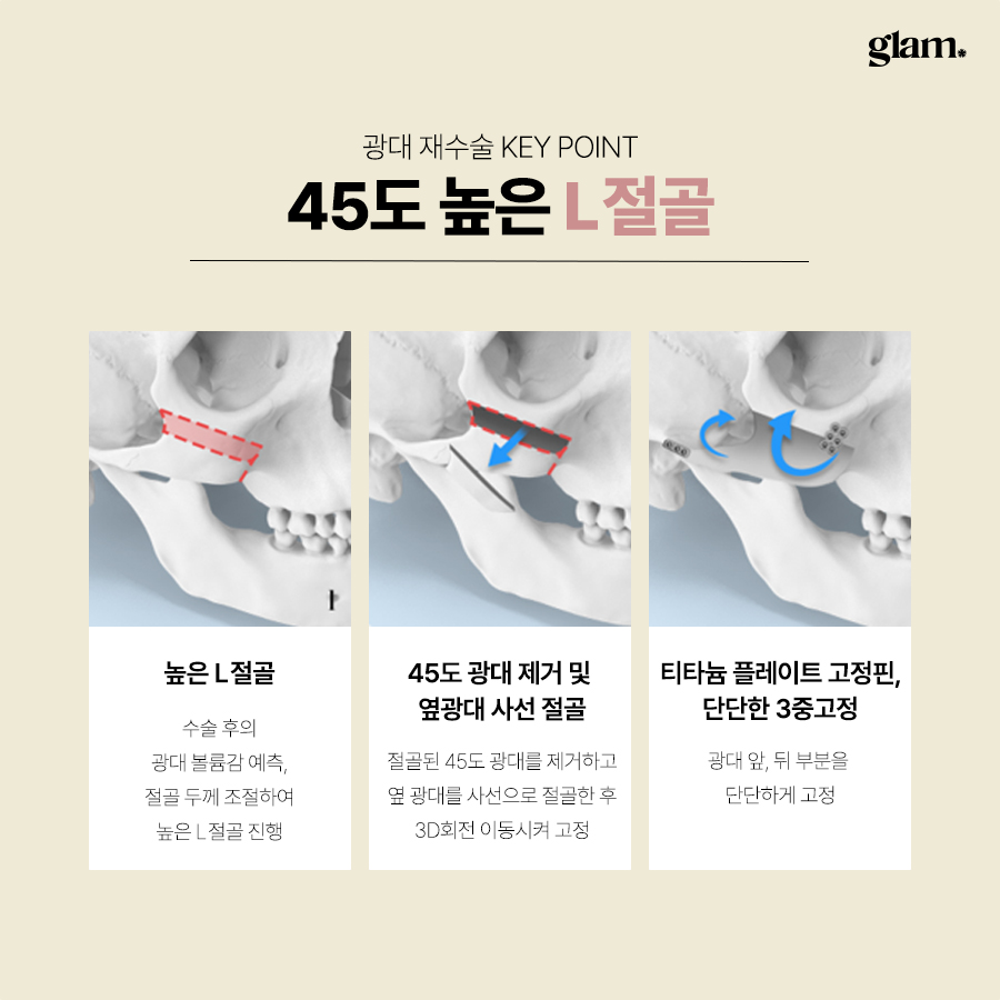
A step deformity is a noticeable “step” or ridge where the zygomatic body and arch meet, or along the previous osteotomy line. Patients often feel this ridge when touching the area, and it can create unnatural shadow lines on the face.
Revision can smooth or recontour the bone surface, adjust fixation, and correct the transition between the zygomatic body and arch. This requires precise three‑dimensional planning to avoid weakening the bone while improving the contour.
Soft tissue sagging after bone reduction
After cheekbone reduction, the soft tissues that used to rest on the bone may have less support. In some people, especially those with heavier or less elastic skin, this can lead to sagging cheeks, deeper nasolabial folds, or more visible jowls.
In revision planning, the surgeon must evaluate not only the bone but also the soft tissues. Sometimes a small additional procedure to support or lift the soft tissue may be recommended together with bone revision, or non‑surgical options may be suggested if bone structure is acceptable.
Functional issues and discomfort
Some patients experience pain around the cheekbone, chewing discomfort, or clicking sensations after their first surgery. These may be related to unstable fixation, malpositioned bone segments, or sinus involvement.
Through imaging and examination, the surgeon checks whether plates and screws are well‑positioned, whether the bone has healed properly, and whether there is any structural cause that revision could reasonably improve.
Preoperative evaluation for revision zygoma surgery
Detailed preoperative evaluation is essential before any revision. The process typically includes:
Imaging
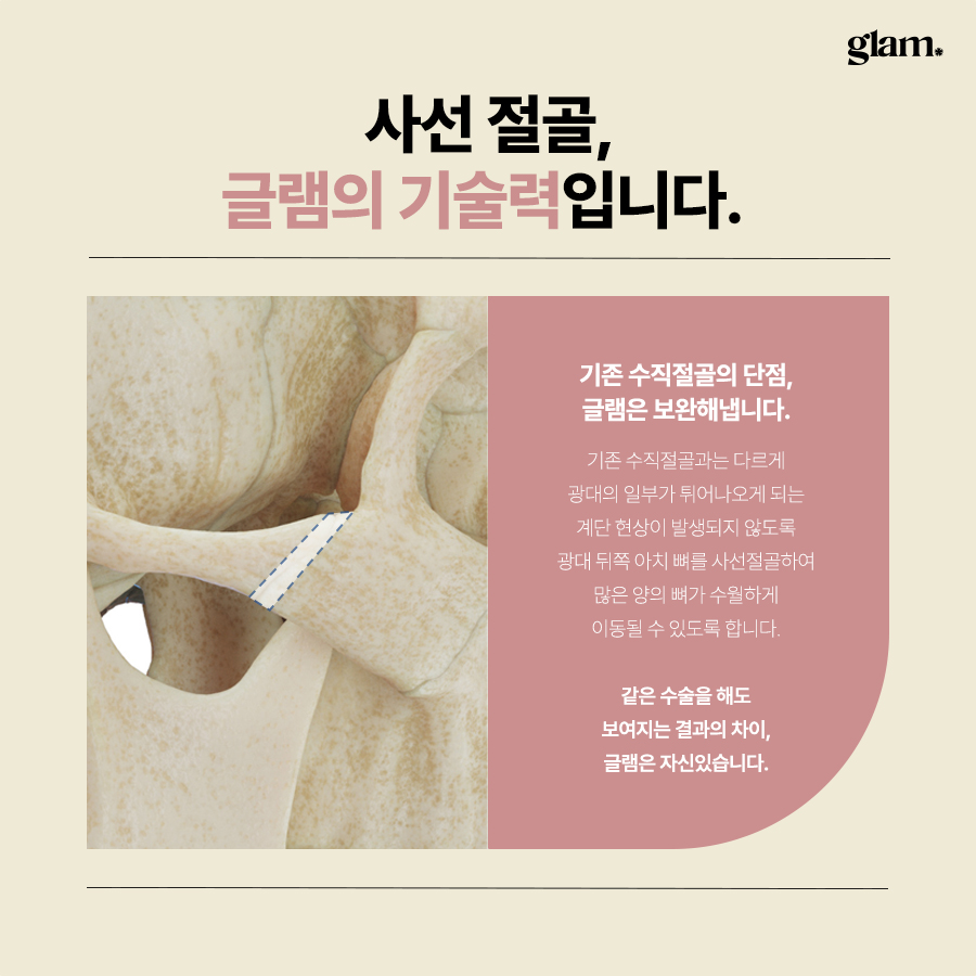
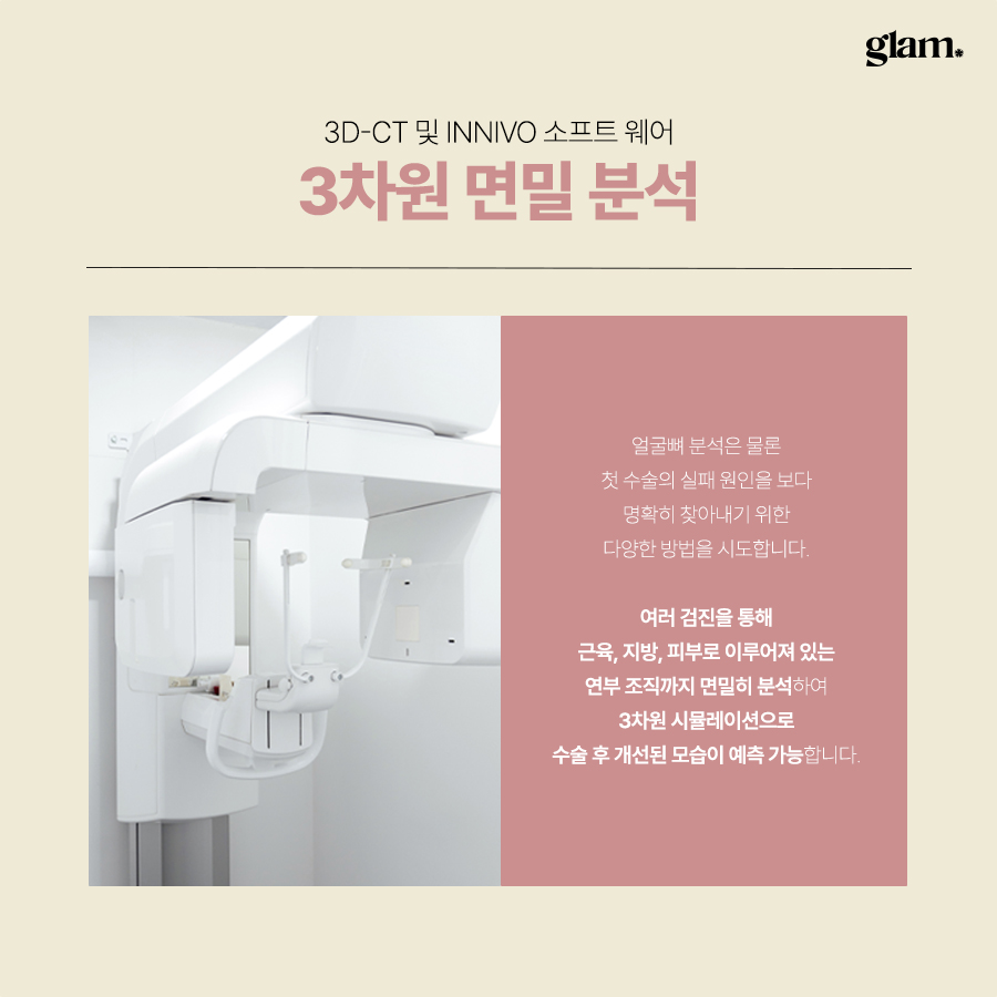
Three‑dimensional CT scans and X‑rays are used to analyze:
Previous osteotomy lines and how the bone was cut.
Current bone position and shape.
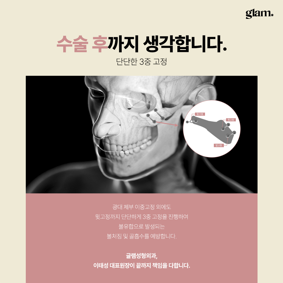
Location and condition of fixation plates and screws.
Bone healing status and any defects.
Sinus involvement or changes.
Soft tissue assessment
The surgeon evaluates:
Skin elasticity and thickness.
Fat volume in the midface and lower face.
Degree of sagging and jowling.
Presence of scar tissue and its effect on mobility.
Facial proportion analysis
The face is assessed from multiple angles: front, 45 degrees, side, and lower view. The goal is to understand how the cheekbones relate to the jawline, nose, chin, and forehead, and how much correction is realistically possible.
Because the clinic focuses only on facial bone surgery, the entire evaluation and planning process is centered on skeletal structure and its relationship with the soft tissue envelope. This helps avoid over‑promising and supports realistic, structural planning.
Surgical approaches and techniques
The exact revision method depends heavily on what was done in the first surgery. During consultation, the surgeon will often ask for previous records or imaging to understand the original technique.
Key concepts include:
Under‑correction
If the cheekbones remain wide, revision may involve additional bone reduction or repositioning. The surgeon may:
Re‑cut the zygomatic body and arch.
Move the bone inward and backward in a more controlled way.
Ensure stronger, more stable fixation to reduce relapse risk.
Over‑correction or sunken midface
When the midface is too flat or sunken, options may include:
Repositioning the zygomatic complex slightly outward or forward.
Using bone grafts or contouring to rebuild deficient areas.
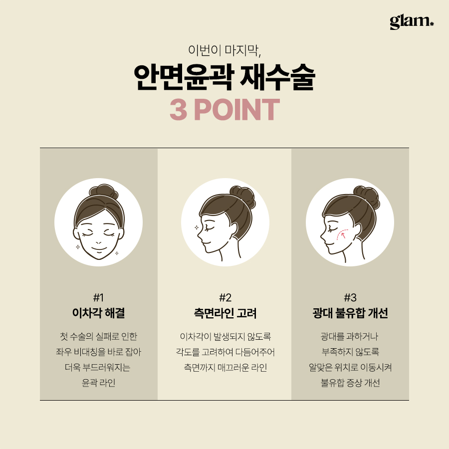
Smoothing irregularities to create more natural curves.
The aim is to restore support and volume while avoiding an over‑projected or artificial look.
Asymmetry correction
For asymmetry, the surgeon may:
Adjust one side more than the other to match height and projection.
Remove or replace plates and screws that are pulling the bone into an unbalanced position.
Combine bone work with soft tissue adjustments if needed.
Step deformities and irregular contours
To correct step deformities, the bone surface can be carefully reshaped. This may involve:
Smoothing sharp edges or ridges.
Re‑aligning segments at the junction of the body and arch.
Re‑fixing segments to create a more continuous contour.
Plates and screws
Existing plates and screws may need to be removed if they are poorly positioned, palpable, or contributing to discomfort. New fixation can then be applied in a more optimal position. Throughout, the surgeon focuses on stable fixation and three‑dimensional planning to support both bone and soft tissue.
Safety, anesthesia, and intraoperative management
Revision zygoma reduction is usually performed under general anesthesia.
Anesthesia plans are tailored to each patient’s health status, surgery length, and expected blood loss. Continuous monitoring of vital signs is maintained throughout the operation. Because revision cases can be longer and more complex than primary surgery, having an in‑house anesthesiologist familiar with facial bone procedures is especially important.
The clinic’s narrow focus on double jaw and facial contouring means the surgical and nursing teams are accustomed to the specific risks and nuances of facial bone revision, including swelling management, nerve protection, and airway considerations.
Recovery, aftercare, and swelling management
Recovery after revision zygoma surgery is similar to primary surgery but may be slightly longer or more unpredictable because of scar tissue and previous changes.
Typical course
Immediately after surgery, patients usually stay in the clinic for monitoring until they are stable. Swelling and bruising are most noticeable during the first week, then gradually decrease over several weeks.
Most patients can return to light daily activities within 1 to 2 weeks, depending on their overall condition and job type. However, the more final facial contour continues to refine over several months as swelling resolves and tissues adapt to the new bone structure.
Supportive aftercare programs
Some clinics may offer adjunctive postoperative care options intended to support recovery; examples include LED light therapy, manual lymphatic drainage, radiofrequency‑based treatments, or other skin‑care modalities. The evidence and expected benefits for these adjuncts vary—ask your treating provider which services are available, which they recommend for your situation, and what outcomes you can reasonably expect.
LED therapy: Used to support skin condition and circulation.
Facial dome treatments: Gentle treatments aimed at promoting blood flow and reducing edema.
Skin reset total care: Programs focused on improving skin texture and comfort after surgery.
Radiofrequency lifting devices: Non‑invasive treatments intended to help with mild skin tightening and swelling control.
These are supportive skin and soft tissue care methods, not substitutes for precise bone surgery. Their role is to help patients feel more comfortable and to support the healing environment after the structural work has been done.
Who may consider revision zygoma surgery?
Revision zygoma surgery may be considered for:
Patients with clear structural problems on imaging, such as malpositioned bone segments, unstable fixation, or significant asymmetry.
Patients with visible deformities like step irregularities, persistent width, or obvious sunken areas.
Patients who experience functional issues, including pain or chewing discomfort, that appear related to bone position or fixation.
Patients with realistic expectations who understand that revision aims for improvement, not perfection or a completely new face.
Patients who have allowed sufficient time for healing after the first surgery. The appropriate timing depends on bone healing and soft tissue condition and should be determined by the surgeon.
Patients without uncontrolled systemic diseases and who can safely undergo general anesthesia.
Online consultation and pre‑visit preparation
For overseas patients, an online consultation is usually the first step. To make this as productive as possible, it helps to prepare the following:
Clear photos of your face from the front, 45‑degree angles, side views, and a lower view looking up.
Any CT scans or X‑rays taken after your first surgery, ideally in DICOM format.
Operation records or a summary from your previous clinic, if available.
A written list of what bothers you most: for example, “right cheek higher,” “step I can feel near ear,” “pain when chewing on left side,” or “face looks too flat from front.”
During online consultation, the surgeon can give a preliminary opinion on whether revision seems structurally possible, what type of imaging will be needed in Korea, and what kind of improvement you may realistically expect. However, final planning always requires in‑person examination and updated imaging.
Before you travel, it is helpful to prepare questions such as:
What are the main structural problems you see in my case?
What type of revision technique are you considering and why?
What level of improvement is realistic, and what will probably not change?
How long should I stay in Korea for surgery and follow‑up?
Expected Korea visit timeline for revision zygoma surgery
Example timeline (varies by case and clinic — confirm specifics with your treating center):
- Initial in‑person consultation and imaging: typically within the first 1–2 days after arrival.
- Surgery and immediate postoperative observation: scheduled per the clinic’s protocol.
- Early postoperative visits and suture removal: timing varies based on the procedure.
- Follow‑up assessments: arranged according to recovery progress.
Recommended length of stay for international patients depends on the procedure, individual recovery, and the clinic’s protocols; confirm the suggested schedule and any travel or recovery recommendations with your clinic before booking.
Day 1–2: In‑person consultation and imaging. You meet the surgeon, undergo CT scans and X‑rays, and have a detailed discussion about the plan, risks, and expected outcome.
Day 3–4: Surgery day. You are admitted, undergo general anesthesia, and have the revision procedure. After surgery, you stay in the clinic for monitoring until stable.
First week: Frequent follow‑ups for wound checks, swelling assessment, and basic aftercare. Some clinics begin gentle supportive treatments such as LED or dome therapy during this period.
Second week: Stitches (if any external ones are used) may be removed, and you can usually prepare for your return flight once the surgeon confirms it is safe.
When planning flights and accommodation, always confirm the recommended duration with the clinic in advance.
Consultation approach and decision‑making
Not every unsatisfactory result requires or is suitable for revision bone surgery. Sometimes, minor asymmetries or mainly soft tissue issues can be better addressed with non‑surgical or less invasive options.
The clinic’s philosophy is to recommend only the minimum necessary surgery. If imaging and examination show that aggressive re‑cutting of bone is not needed, the surgeon will explain alternative approaches, such as soft tissue procedures or non‑surgical treatments.
During consultation, there should be a thorough, honest discussion about:
The exact structural findings on imaging.
Which issues revision can realistically improve.
Which concerns are more related to soft tissue or personal perception.
Surgical risks, possible complications, and limitations.
Recovery timeline and follow‑up requirements.
For overseas patients, it is particularly important to understand how communication and care will be handled after you return home. Ask how to share post‑operative photos, how often to check in, and what to do if you have concerns later.
Frequently asked questions
Can revision zygoma surgery completely fix my first surgery result?
In most cases, revision aims for noticeable improvement, not complete reversal or perfection. Because the bone has already been cut and healed in a new position, there are anatomical limits to how much it can be changed again. Scar tissue and previous bone removal also restrict options. A careful surgeon will explain what is realistically achievable in your specific case, focusing on structural balance and function rather than dramatic transformation.
How long should I wait after my first zygoma surgery before considering revision?

The ideal timing depends on how your bone and soft tissues have healed. In general, surgeons prefer to wait until bone healing is solid and most swelling has resolved, which often means several months or longer. In some situations, if there is a clear structural problem such as unstable fixation, earlier intervention may be considered. The surgeon will determine appropriate timing after reviewing imaging and examining you.
Is revision zygoma surgery more risky than the first surgery?
Revision is usually more complex because of previous bone cuts, plates and screws, and scar tissue. This can increase technical difficulty and sometimes the risk of certain complications. However, with detailed imaging, careful planning, and an experienced facial bone surgeon working with a dedicated anesthesiologist, many risks can be managed. It is important to have a clear discussion about potential complications and how the clinic prepares for them.
How do I know if I really need bone revision or just soft tissue treatment?
Imaging is the key. If CT scans show that the bone position and fixation are stable and well‑aligned, but your concerns are mainly about mild sagging or small asymmetries, soft tissue treatments may be more appropriate. If imaging reveals malpositioned bone, step deformities, or unstable fixation, then bone revision may be considered. During consultation, the surgeon should explain whether your main issues are skeletal, soft tissue, or both, and recommend the least invasive option that addresses the true cause.
Reassurance and next steps
Feeling disappointed or anxious after a previous zygoma reduction is very common, especially when you see asymmetry, irregular lines, or changes you did not expect. It is understandable to be cautious about another surgery.
With a surgeon experienced in facial bone revision and a clinic system dedicated to facial skeletal procedures, it is often possible to achieve a more balanced, natural‑looking result and improved comfort. The key is careful analysis, realistic goal‑setting, and a conservative, safety‑first approach.
If you are considering revision zygoma surgery in Seoul, an online consultation can be a good first step. By sharing your photos, imaging, and concerns in detail, you can receive a more personalized assessment and decide calmly whether traveling for revision is the right choice for you.






댓글
댓글 쓰기