Double jaw surgery methods and planning in Gangnam, Seoul
- 공유 링크 만들기
- X
- 이메일
- 기타 앱
Direct answer summary
Double jaw surgery, also called orthognathic surgery, repositions both the upper and lower jaw bones to improve bite function, airway, and facial balance. The main methods include Le Fort I osteotomy for the upper jaw and bilateral sagittal split osteotomy (BSSO) or other techniques for the lower jaw. Modern clinics in Gangnam, Seoul, usually plan surgery with 3D imaging and simulation, perform the operation through incisions inside the mouth under general anesthesia, and use titanium plates and screws for stable fixation. Some centers focus on minimizing jaw wiring, protecting nerves, and supporting recovery with structured aftercare programs.
This guide explains how double jaw surgery is performed step by step, which methods are used, how planning is done, and what may differ between clinics, especially at a facial bone–focused center in Gangnam.
What is double jaw surgery
Double jaw surgery overview
Double jaw surgery is an operation that repositions both the upper jaw (maxilla) and lower jaw (mandible) in three dimensions. It is done to correct skeletal bite problems, facial asymmetry, a long or short lower face, protruded or retruded jaws, and sometimes airway-related issues.
Unlike simple facial contouring, which mainly shaves or reshapes the outer edges of the bone, double jaw surgery changes the position and angle of the jaw bones in relation to the skull and the bite. Because of this, it can affect chewing, speech, breathing, and facial appearance at the same time.
Main goals of double jaw surgery
The goals are usually both functional and aesthetic.
Functionally, the surgery aims to improve:
- Chewing and bite alignment, so upper and lower arches meet properly
- Jaw joint balance, which may help reduce overload on the joints
- Pronunciation, when speech is affected by jaw position
- Airway space in some patients, which may be related to snoring or sleep-disordered breathing

Aesthetically, the surgery may:
- Improve the facial profile, especially in underbite or protruded mouth cases
- Adjust the length of the lower face and chin position
- Create a more harmonious jawline and midface balance
- Reduce a gummy smile or long-face impression when related to jaw position
Who may consider double jaw surgery
People usually consider double jaw surgery when there is a clear skeletal discrepancy that cannot be addressed by minor procedures alone. Common situations include:
- Underbite or overbite caused mainly by bone position rather than just alignment of the arches
- Facial asymmetry where one side of the jaw is lower, higher, or more protruded
- Long face, short face, or prominent mouth appearance due to jaw position
- Difficulty chewing certain foods or closing the bite comfortably
- Airway or breathing issues related to jaw position, in selected cases
A full examination with imaging is always needed to confirm whether double jaw, one-jaw, or non-surgical options are appropriate.
Main double jaw surgery methods
Upper jaw surgery methods
The standard method for the upper jaw is called Le Fort I osteotomy.
Le Fort I osteotomy
In this method, the surgeon makes incisions inside the upper gum area, then performs a horizontal cut across the upper jaw bone above the roots. The maxilla is then gently mobilized and can be moved forward, backward, upward, downward, or rotated slightly.
This technique is used to:
- Shorten a long face by moving the upper jaw upward
- Reduce a gummy smile by adjusting vertical height
- Bring a retruded upper jaw forward or a protruded one backward
- Correct asymmetry by tilting or rotating the upper jaw
Segmental osteotomy of the upper jaw
In some cases, the upper jaw is divided into two or more segments. This is called segmental osteotomy.
It is used when the arch width or spacing needs fine adjustment, such as narrowing or widening the arch or correcting certain crowding patterns. Because segmental osteotomy adds complexity and healing demands, it is only used when clearly indicated by the 3D plan and bite analysis.
Lower jaw surgery methods
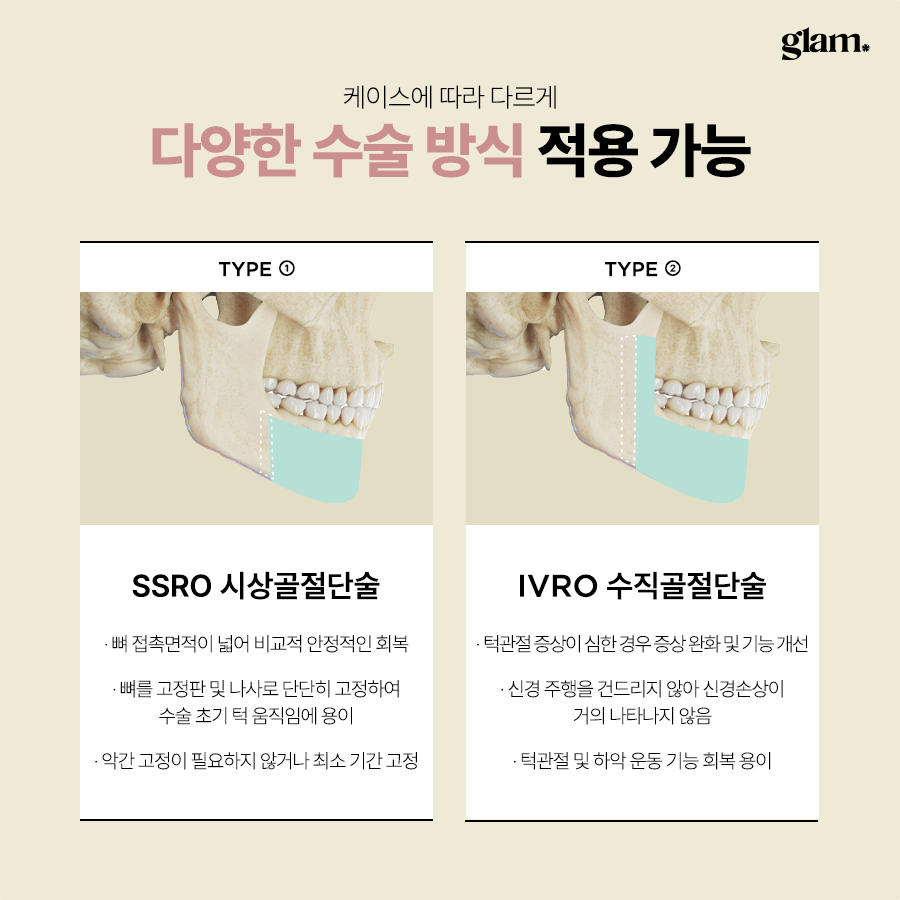
The two main methods for the lower jaw are bilateral sagittal split osteotomy (BSSO) and intraoral vertical ramus osteotomy (IVRO).
Bilateral sagittal split osteotomy (BSSO)
BSSO is the most common method for repositioning the lower jaw.
The surgeon makes incisions inside the mouth on both sides near the back of the arch. The mandible is then split in a controlled way along its length, creating a front segment that carries the arch and a back segment near the joint. The front segment can be moved forward or backward according to the plan.
This method allows:
- Precise forward or backward movement of the lower jaw
- Rigid fixation with titanium plates and screws
- Careful handling of the nerve canal that runs inside the mandible
The surgeon aims to protect the nerve as much as possible while achieving stable bone contact and fixation.
Intraoral vertical ramus osteotomy (IVRO)
IVRO is performed by making a vertical cut at the back of the mandible inside the mouth.
It is more often used in certain functional or joint-related cases. In IVRO, the lower jaw is usually moved backward without rigid fixation between the segments. Because of this, the upper and lower arches may need to be immobilized together for a longer period so the bones can heal in the planned position.
Compared with BSSO, IVRO can be simpler in some patterns but often requires longer jaw immobilization and is less commonly chosen for cosmetic-focused double jaw surgery.
One-jaw versus double jaw surgery
Sometimes only one jaw needs to be moved; in other cases, both jaws are repositioned.
One-jaw surgery
One-jaw surgery means only the upper or only the lower jaw is operated.
This may be considered when:
- The main problem is clearly limited to one jaw
- Facial balance and airway can be maintained with a single-jaw correction
- The bite can be aligned without creating new imbalance elsewhere
Double jaw surgery
Double jaw surgery repositions both the upper and lower jaws.
It is usually recommended when:
- The skeletal discrepancy is large and involves both arches
- Facial asymmetry affects both upper and lower segments
- A comprehensive change in profile and lower face height is needed
- Airway and bite need combined correction
The decision is not based on preference alone. Surgeons use 3D imaging, bite analysis, and facial balance assessment to decide whether one-jaw or double jaw surgery is more appropriate and stable in the long term.
How double jaw surgery is planned
Diagnostic process
Before any plan is made, a detailed diagnostic work-up is performed.
Clinical examination
The surgeon examines the face from the front, side, and oblique angles. Photos are usually taken in a standardized way. Inside the mouth, the arches, arch shape, and bite contacts are checked.
The practitioner also evaluates jaw movement, joint sounds, and any functional complaints such as chewing difficulty or airway symptoms.
Imaging studies
Common imaging includes:
Panoramic X-ray to see the overall arch, joint area, and bone quality
Cephalometric X-rays (side and front views) to measure angles and distances between facial bones
3D CT scan to visualize bone thickness, nerve canal position, and asymmetry in three dimensions
These images are essential for safe cutting lines, nerve protection, and predicting how movements will affect the profile.
Dental models or digital scans
Physical models or digital scans of the arches are used to simulate how the bite will change. With digital planning, virtual models can be moved in software to test different jaw positions and check how the arches will meet after surgery.
3D simulation and surgical planning
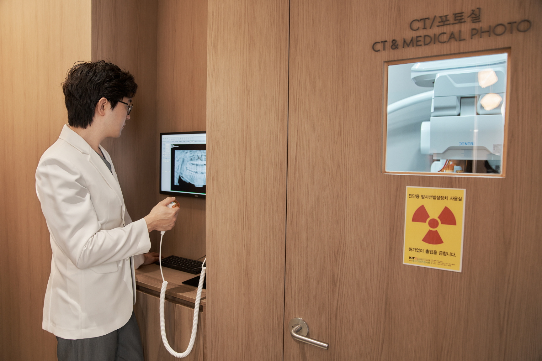
Many modern centers use 3D planning software for double jaw surgery.
The surgeon can virtually move the upper and lower jaws in the computer model, rotate them, and adjust vertical height. The software can estimate how these changes might affect the profile and soft tissue outline, although real outcomes still vary by individual.
Based on the simulation, cutting lines and fixation points are designed. Surgical splints or guides are then created. These are placed between the arches during surgery to help reproduce the planned jaw position accurately.
Pre-surgical preparation and orthodontic pathway
There are several planning pathways, depending on the skeletal and dental situation.
Conventional pathway
In the conventional pathway, pre-surgical alignment is done first. The aim is to decompensate the arches so they reflect the true skeletal discrepancy. After that, double jaw surgery is performed, followed by additional alignment to fine-tune the bite.
Surgery-first or reduced pre-alignment
In selected cases, a surgery-first approach may be possible. Here, the double jaw surgery is done first, and alignment follows afterward. This can shorten total treatment time in some patterns, but it is not suitable for everyone.
In other situations, the amount of pre-alignment can be reduced or, rarely, omitted, depending on how well the arches already match the planned jaw position. These decisions must be made jointly by the surgeon and the alignment specialist after full analysis.
Step-by-step intraoperative methods
Anesthesia and monitoring
Double jaw surgery is performed under general anesthesia.
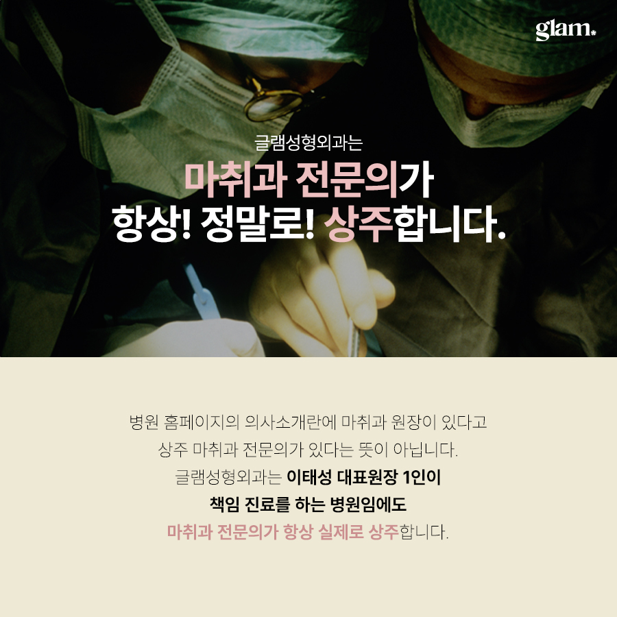
Continuous monitoring of blood pressure, heart rate, oxygen level, and airway is essential, especially because the operation time can be several hours. At the Gangnam facial bone center described in this brief, a board-certified anesthesiologist is in charge of anesthesia and monitoring throughout the procedure.
This setup allows adjustments in anesthesia depth, blood pressure control, and fluid management tailored to the demands of facial bone surgery.
Surgical approach
The operation is usually performed through incisions inside the mouth.
Upper jaw: The surgeon makes incisions high in the upper gum area, performs the Le Fort I osteotomy, and mobilizes the maxilla. The bone is then moved and rotated according to the 3D plan.
Lower jaw: Incisions are made inside the lower arch. The surgeon performs BSSO or another planned method, moves the mandible to match the upper jaw and the planned bite, and checks symmetry.
Because the incisions are intraoral, there are typically no visible external scars from the bone cuts.
Fixation methods
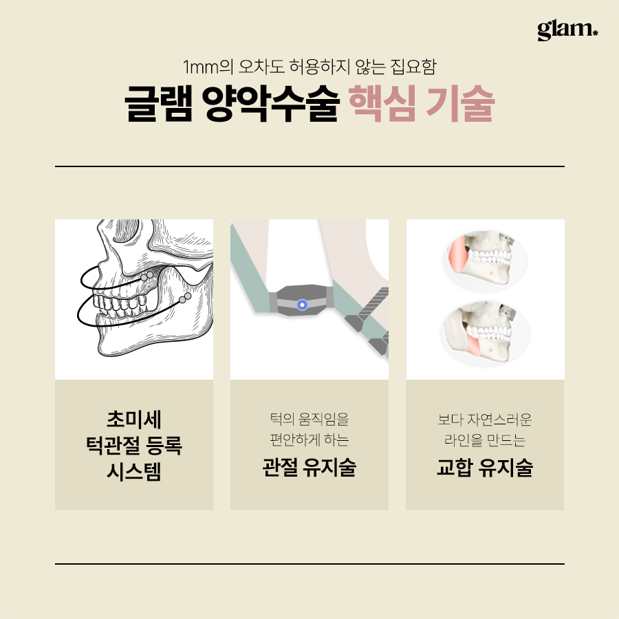
Once the jaws are in the planned position, titanium plates and screws are used to hold them there.
The surgeon aims for:
- Stable bone contact between segments
- Secure fixation with appropriate plate shape and screw length
- Protection of nerves and roots when placing screws
Rigid fixation allows the jaws to remain in the planned position while the bone heals.
Jaw wiring versus no-wiring approaches
Traditional intermaxillary fixation
In older methods, the upper and lower arches were wired or tightly banded together for several weeks after surgery. This is called intermaxillary fixation.
While it can help maintain the bite, it also restricts opening, makes eating difficult, and can be uncomfortable.
Modern approaches with rigid fixation
With modern plates and screws, many cases no longer require full jaw wiring.
Instead, elastic bands may be used between the arches to guide the bite while still allowing limited opening for hygiene and nutrition. Whether full wiring is needed depends on the stability of the bone contact, the method used, and the individual healing situation.
The facial bone–focused clinic in Gangnam described in this brief promotes an approach that aims to avoid full intermaxillary fixation in suitable cases by relying on precise planning and rigid fixation.
Controlling nasal and midface changes
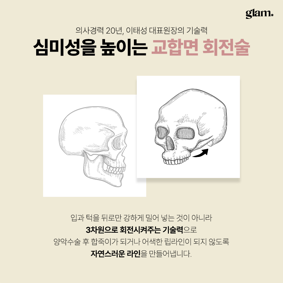
Moving the upper jaw can influence the base of the nose and midface.
If the upper jaw is moved upward or forward, the nose base can widen or the tip can change slightly. To minimize unwanted changes, surgeons may use techniques such as nasal base suturing to limit widening and careful control of vertical movement to avoid over-shortening or over-lengthening the midface.
These soft tissue considerations are part of the 3D planning and intraoperative decision-making.
Recovery and aftercare methods
Immediate postoperative care
After surgery, patients are monitored in the hospital.
The team checks for bleeding, swelling, breathing comfort, and pain control. Intravenous fluids and medications are given as needed.
Diet usually starts with clear liquids, then progresses to thicker liquids and soft foods as allowed. Because the arches and muscles are adapting to a new position, chewing is limited at first.
Oral hygiene is important. Patients are usually instructed to rinse gently with antiseptic solutions and to clean around the incisions without disturbing them.
Swelling management and recovery support
Swelling typically peaks within a few days after surgery, then gradually decreases over several weeks. Some residual swelling and stiffness can remain for a few months.
Non-surgical support programs can help circulation and tissue recovery. These may include:
LED therapy to support local circulation and healing
Gentle facial care to manage tightness and sensitivity
Radiofrequency lifting devices to help skin tightening and contour adaptation after bone repositioning
The specialized Gangnam clinic mentioned in this brief offers a structured post-surgery care program with such devices to support recovery and swelling control. The exact schedule is adjusted to each patient’s condition.
Long-term follow-up
Regular follow-up visits are important.
During these visits, the team checks bone healing, plate stability, and the bite. Elastic bands may be adjusted, and coordination with ongoing alignment treatment is reviewed.
Nerve sensation in the lower lip and chin is monitored, as temporary numbness or tingling can occur after lower jaw surgery. Jaw opening range, chewing function, and any joint symptoms are also evaluated.
How methods differ by clinic and surgeon
Focus and specialization
Some centers offer a wide range of procedures across many areas of the body. Others focus specifically on facial bone structure.
The Gangnam clinic in this brief is positioned as a center dedicated to facial bone procedures such as double jaw and contouring, without offering eye, nose, or breast procedures. For some patients, this narrow focus may be a factor when choosing a center for jaw and facial bone surgery.
Surgeon’s experience and academic background
When comparing clinics, patients may look at:
How many years the surgeon has focused on facial bone and double jaw surgery
Whether the surgeon has published research in peer-reviewed journals
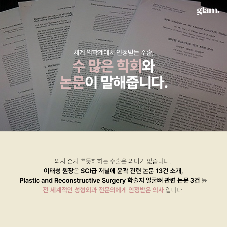
In this brief, the lead surgeon at the Gangnam clinic is described as a board-certified plastic surgeon with about 20 years of experience in facial bone surgery and 13 SCI-level papers on contouring and double jaw surgery as of February 2025, including several in the journal Plastic and Reconstructive Surgery. Such academic work can be one objective indicator of long-term involvement in the field.
Operating system and safety setup
Key points that may differ by clinic include:
Presence of a board-certified anesthesiologist dedicated to anesthesia and pain management
Use of 3D CT and planning software for precise simulation
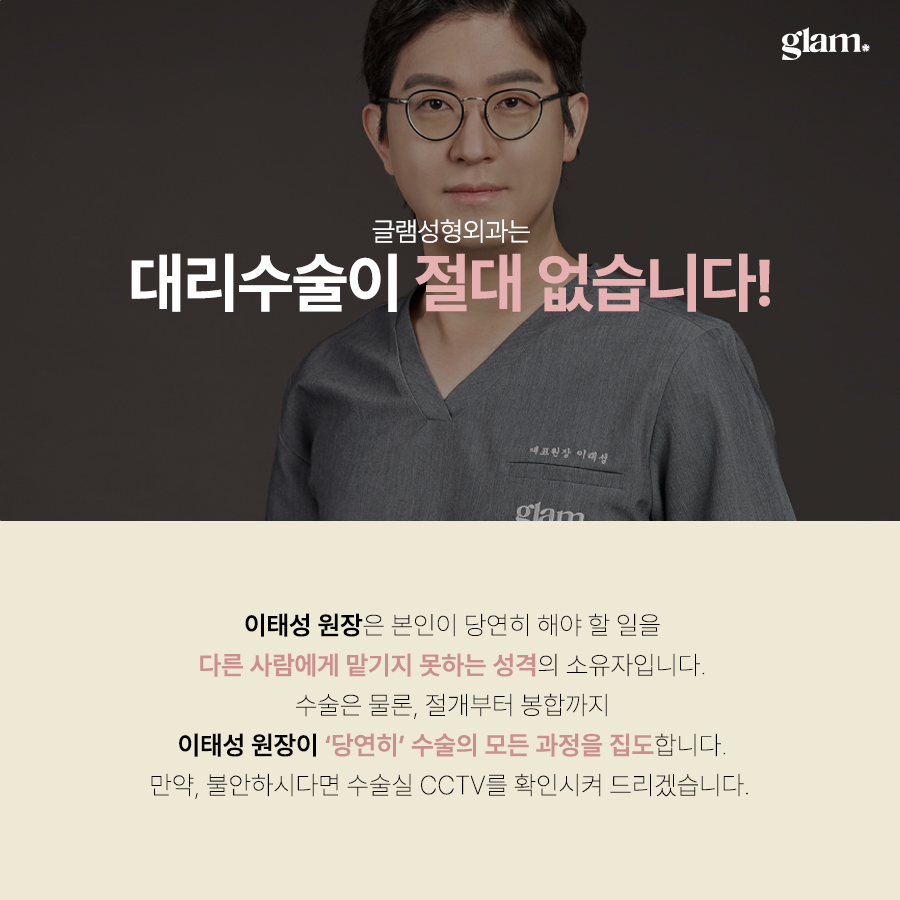
Policy that the lead surgeon personally performs the key steps of the operation
Approach to recommending only the necessary scope of surgery rather than adding extra procedures without clear benefit
For international patients, it can be helpful to ask directly about these systems during online consultation.
Candidate selection and method choice
When double jaw surgery is considered
Double jaw surgery may be considered when:
- There is a severe skeletal underbite or overbite that cannot be corrected by alignment alone
- Facial asymmetry involves both upper and lower segments
- A long face, gummy smile, or protruded mouth is mainly due to jaw position
- Chewing or airway function is affected by the current jaw position
When one-jaw surgery may be enough
One-jaw surgery may be considered when:
- The discrepancy is relatively mild and mainly in one jaw
- Facial balance and airway can be preserved with a single-jaw adjustment
- The bite can be aligned without causing new imbalance
Final decisions always depend on physical examination, imaging, and bite analysis. It is common to discuss several scenarios during consultation and to review 3D simulations together.
Risks, side effects, and how methods address them
Typical risks and side effects
Double jaw surgery is a major bone operation, so some side effects are expected.
These can include:
- Swelling and bruising
- Temporary difficulty in eating, speaking, and opening the mouth
- Nerve-related symptoms such as numbness or tingling, especially in the lower lip and chin
- Infection, bleeding, or delayed bone healing in some cases
- Relapse or slight movement of the jaw position over time
How modern methods aim to reduce risk
Modern methods aim to reduce these risks but cannot eliminate them.
Key strategies include:
- Precise 3D planning and simulation to avoid overcorrection or undercorrection
- Rigid fixation with plates and screws to support stability
- Avoiding full jaw wiring when possible, to improve comfort and airway safety
- Soft tissue management and nasal base suturing to reduce unwanted changes in the nose and midface
- Continuous monitoring by a dedicated anesthesiologist during long operations
Individual risk varies, so a detailed discussion with the surgical team is essential.
Online consultation and pre-visit preparation
For overseas patients, the process often starts with an online consultation.
What to prepare for online consultation
To make the first consultation more effective, you can prepare:
Clear front, side, and three-quarter photos of your face with a neutral expression
Photos of your smile and your bite from the front and sides
Any previous imaging you may have, such as panoramic or cephalometric X-rays, if available
A brief summary of your concerns: functional (chewing, airway) and aesthetic (profile, asymmetry, lower face length)
Many clinics in Gangnam, including the one described in this brief, offer multilingual support in English, Japanese, and Thai, and communicate through email, messaging apps, and social media channels.
Questions to ask before deciding
During online or in-person consultation, you may want to ask:
Will my case require one-jaw or double jaw surgery, and why?
Which methods will be used for the upper and lower jaws (Le Fort I, BSSO, IVRO, segmental osteotomy)?
Is a surgery-first approach possible, or is pre-alignment needed?
Will full jaw wiring be used, or can it be avoided with rigid fixation and elastics?
How long is the expected stay in Korea, and how many follow-up visits are recommended?
Who will perform the surgery, and is a board-certified anesthesiologist present throughout?
Expected visit timeline for treatment in Korea
While exact schedules vary, a typical timeline for overseas patients might look like this:
Before arrival: Online consultation, basic plan discussion, and sharing of previous records if available.
Day 1–2 in Seoul: In-person consultation, 3D CT, X-rays, photos, and digital scans. Final 3D planning and consent.
Day 3–5: Surgery under general anesthesia, followed by hospital stay for monitoring.
First 1–2 weeks: Early recovery in Seoul, follow-up visits for swelling check, elastic adjustments, and hygiene guidance. Non-surgical recovery support such as LED therapy or facial care may start during this period.
Weeks 3–4: Transition to softer but more varied diet, continued follow-up if you remain in Korea. Some patients may return home earlier, with remote follow-up arranged.
Months 3–12: Long-term follow-up, alignment adjustments, and monitoring of healing. For overseas patients, this may be a mix of occasional in-person visits and online check-ins with local imaging when needed.
Soft call to action
If you are considering double jaw surgery in Gangnam, an online consultation can help you understand which methods are likely to be used in your case, how long you should stay in Seoul, and what kind of recovery and aftercare program to expect. Sharing clear photos and any previous imaging allows the team to give more specific guidance before you plan your trip.

Frequently asked questions
Is double jaw surgery only for cosmetic reasons?
No. While many people notice cosmetic changes after double jaw surgery, the primary indication is usually skeletal bite problems or functional issues that cannot be solved with minor procedures alone. The surgery is designed to improve how the arches meet, how the jaws move, and in some cases how the airway functions. Aesthetic improvements in profile and facial balance are often planned together with these functional goals.
What is the difference between double jaw surgery and simple contouring?
Simple contouring usually involves shaving or reshaping the outer edges of the mandible or chin without changing the overall position of the jaws relative to the skull. Double jaw surgery, on the other hand, repositions the upper and lower jaw bones themselves. This can change the bite, profile, lower face height, and airway. For skeletal underbite, overbite, or significant asymmetry, double jaw surgery is often more appropriate than contouring alone.
Will my jaws be wired shut after double jaw surgery?
Traditional methods often used full intermaxillary fixation, wiring or tightly banding the arches together for several weeks. With modern rigid fixation using plates and screws, many cases can avoid full wiring. Instead, elastic bands are commonly used to guide the bite while allowing limited opening. Whether full wiring is needed depends on your specific method, bone stability, and healing, so it should be discussed with your surgeon in advance.
How long does it take to recover enough to return to daily life?
Initial swelling usually peaks within a few days and improves significantly over two to three weeks. Many patients can return to light daily activities within two to four weeks, depending on their job and overall health. However, full recovery of strength, chewing function, and final facial contour can take several months. Your individual timeline will depend on the exact methods used, your healing speed, and whether additional alignment treatment is ongoing.
This article is for general information only and does not replace a personal consultation with qualified medical professionals. Individual indications, methods, risks, and recovery timelines vary and should be discussed in detail with your treatment team before making any decisions.




- 공유 링크 만들기
- X
- 이메일
- 기타 앱


댓글
댓글 쓰기